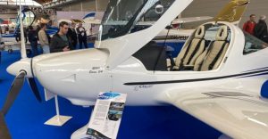
• Applications
    • All Applications
    • Non-Destructive Testing
    • Material Analysis
    • Biomedical
    • Space
    • Education
    • Others
  • Products
    • All Products
    • MiniPIX series
      • MiniPIX SPRINTER
      • MiniPIX TPX3
      • MiniPIX TPX Standard
      • MiniPIX SPACE
      • MiniPIX EDU
      • MiniPIX TPX3 Flex
    • WidePIX series
      • WidePIX L 2(1)x5
      • WidePIX L 2(1)x10
      • WidePIX L 2(1)x15
      • WidePIX 5×5 MPX3
    • AdvaPIX
    • EDU Kit
    • Custom products
    • Software
      • PIXet Pro
      • Pixet Basic
      • TraX Engine
  • Technology
  • News
  • Company & Career
  • Science
    • Science department
    • Research Projects
    • Publications
  • Support
    • Downloads
    • Tutorials & FAQ
    • SW Documentation
  • Contact
Contact

News

Filters
Reset
In this image, we showcase NASA astronaut Katherine Megan McArthur aboard the International Space Station (ISS). Highlighted in a red circle at the top of the image is our MiniPIX TimePIX Radiation Monitoring Camera, a key tool designed to safeguard the astronauts and the sensitive electronic and computer systems onboard. Adjacent to the MiniPIX, you will spot a golden device; this is the original, comparatively larger, and less efficient radiation monitor of the ISS. Centre stage, circled in red, stands the robust HERA Radiation Monitor - a product of NASA's ingenuity, equipped with ADVACAM's Single-photon counting chip. Image courtesy of NASA
Applications
03/14/2023

SPACE WEATHER MONITORING

Applications: Space Charged particles from solar flares and coronal ejections…
Read more ›
The picture shows an experimental Flash proton beam measurement at OncoRay Clinique in Dresden. A so-called „water phantom“ (a box filled with water) equipped with a remotely controlled positioning system is used for calibration before each treatment day in the facility. Advacam used the phantom to characterize stray radiation of Flash proton beam by a specifically modified detector, based on MiniPIX Timepix3 Flex architecture equipped with a silicon sensor.
Applications
03/14/2023

MINIMIZING SIDE EFFECTS OF INNOVATIVE CANCER RADIOTHERAPY

Applications: Biomedical Our single-photon counting detectors participated in a groundbreaking…
Read more ›
Applications
03/14/2023

FOOD INSPECTION: X-RAY CLASSIFICATION AND QUALITY CONTROL

Applications: Biomedical Our single-photon-counting cameras allow for the creation of…
Read more ›
This image shows a slow-motion recording of a mouse's heartbeat. The rodent's heart beats at a rate of 670 beats per minute, ten times faster than a human heart. The recording was made with our AdvaPIX detector at a rate of 1000 frames per second. Bone tissue was suppressed using post-processing to make the heart visible even behind the rib cage. In the video, you can observe how the individual chambers and atria of the heart darken or lighten, which is the actual movement of blood inside the organ. In this case, no contrast agent was used.
Applications
03/14/2023

FUNCTIONAL REAL-TIME TISSUE IMAGING

Applications: Biomedial Our cameras provide real-time, high-speed X-ray videos of…
Read more ›
There are two outputs of our single-particle camera. On the left, there's an image showcasing anatomic features: The veins or arteries of mouse lungs clearly. The right image employs a detector to visualize a specific micro-structure: The lung's alveoli, allowing for the observation of their distribution within the organ anatomy. The X-ray imaging system utilized in this study merges a microfocus X-ray tube with a large-area photon counting detector, WidePIX 5X10, having a resolution of 2560 x 1280 pixels with a pitch of 55 µm.
Applications
03/14/2023

SOFT TISSUES DIFFERENTIATION

Applications: Biomedical Our innovative imaging cameras employ advanced technology for…
Read more ›
Spectral computed tomography slices where each colour represents a tissue type. Picture taken by AdvaPIX
Applications
03/14/2023

SPECTRAL COMPUTED TOMOGRAPHY

Spectral computed tomography enhances 3D imaging, advancing tissue identification and…
Read more ›
ADVACAM SPECTRAL RADIOGRAPHY: Material discriminating spectral imaging of a mouse. Colors represent different tissue types.
Applications
03/14/2023

SPECTRAL RADIOGRAPHY

Applications: Biomedical Photon counting detectors excel in bio-imaging due to…
Read more ›
News
03/14/2023

Exploring Innovations in Particle Detection: Highlights from the Medipix Collaboration Meeting

At the recent Medipix Collaboration Meeting, we saw some incredible…
Read more ›
Applications
03/13/2023

MORE PRECISE HEAD CANCER TUMOR IRRADIATION

Applications: Biomedical ADVACAM detectors are being tested as a method…
Read more ›
Applications
03/13/2023

ROBOT MONITORS RADIOACTIVE DRUGS IN THE BODY

Applications: Biomedical A newly developed robotic device called ThyroPIX could…
Read more ›
Examples of measurements with Timepix detectors utilized by our spin-off company AdvaScope. On the left side: An EBSD diffraction pattern of Nickel. On the right: A diffraction pattern measured for the Si sample aligned to [100] zone axis. Below: An example of an implementation of a Timepix3 detector together with the retraction device that has been installed on a TEM. The external electronics are located and attached to the casing. It is connected via a PCB feedthrough to the vacuum chamber.
Applications
03/13/2023

NOISELESS AND FAST ELECTRON MICROSCOPY

Applications: Material Analysis Our single-photon counting detectors provide a highly…
Read more ›
ADVACAM Ore quality identification: Multiple X-ray transmission images are taken with different energy sensitivity (threshold). Each transmission scan is converted to an equivalent thickness of reference material (e.g., aluminum). Different materials in a sample show different spectral dependencies. If the actual imaged material equals the reference one, then both images show the same equivalent thickness.
Applications
03/13/2023

REVOLUTIONARY ORE QUALITY IDENTIFICATION

Applications: Material Analysis Our energy-sensitive spectral X-ray cameras facilitate color…
Read more ›
Applications
03/13/2023

MATERIAL-SENSITIVE X-RAY INSPECTION

Applications: Material Analysis Our energy-sensitive X-ray cameras enable color radiography…
Read more ›
ADVACAM Material Analysis: XRD MINERAL MAPPING - examined 5mm thick peace of mineral. The diffractograms in this image visualize different materials inside the examined 5mm thick peace of mineral. The AdvaPIX TPX3 CdTe camera was used to make the measurements.
Applications
03/13/2023

XRD MINERAL MAPPING

Applications: Material Analysis Imagine the ability to create detailed maps…
Read more ›
ADVACAM: - XRD IN-DEPTH QUENCHING AND WELD INSPECTION: Partially quenched steel sample: The quenching level was measured in 7 points, as shown in the graph. At every point, the level of quenching can be determined. The image was done by AdvaPIX CdTe.
Applications
03/13/2023

XRD IN-DEPTH QUENCHING AND WELD INSPECTION

Applications: Material Analysis Quenching alters the inner structure of a…
Read more ›
ADVACAM Material Sensitive X-ray fluorescence
Applications
03/13/2023

X-RAY FLUORESCENCE: DISTINGUISH ELEMENT COMPOSITION INSTANTLY

Applications: Material Analysis Achieve speed, precision and accuracy with full…
Read more ›
The XRD measurements performed in transmission mode (X-ray beam penetrates through the sample, detector is placed behind of it) with polychromatic X-ray beam of 160 kVp. Anisotropic scattering patterns indicate presence of texture in the sample structure. Color coding in these images shows powder signal in red, reflections from larger crystals in green and mean energy in blue. An AdvaPIX camera was used to create this image.
Applications
03/13/2023

X-RAY DIFFRACTION AND CRYSTALLOGRAPHY

Applications: Material Analysis X-ray diffraction is an analytical method based…
Read more ›
ADVACAM Non-destructive testing: SPECTRAL IMAGING FOR NON-DESTRUCTIVE TESTING. An example of spectral X-ray imaging is the differentiation of materials in electronics. A laptop was scanned using the Widepix MPX3 CdTe detector on the robotic scanner. The resulting scan has 7852x9880 pixels, i.e., 77.6M pixels. Both the grayscale and “color” X-ray images are shown.
Applications
03/13/2023

SPECTRAL IMAGING FOR NON-DESTRUCTIVE TESTING

Applications: NDT, non-destructive testing Color X-ray imaging is now achievable.…
Read more ›
ADVACAM Non-destructive testing: INSPECT COMPOSITE FIBER ORIENTATION. The orientation of fibers in a composite material is clearly visualized at a resolution of 55 µm. In the upper image, you can see light aircraft composite part. It shows fiber bunches' alignment, distribution, and location in high resolution. Below is a picture of a sports car composite door where individual fibers can be detected.
Applications
03/13/2023

INSPECT COMPOSITE FIBER ORIENTATION

Applications: NDT, non-destructive testing Inspect composite fiber orientation using low-energy…
Read more ›
ADVACAM Non-destructive testing: UNCOVER COMPOSITE DELAMINATION. This picture shows aluminum core delaminations in a honeycomb composite part consisting of the metallic core and a CFRP skin. Sharper edges signal the absence of glue. Some bubbles in epoxy and Carbon fiber bunches are visible as well.
Applications
03/12/2023

UNCOVER COMPOSITE DELAMINATION

Applications: NDT, non-destructive testing Advacam produces novel photon-counting x-ray imaging…
Read more ›
ADVACAM Non-destructive testing: LIGHT COMPOSITE INSPECTION. An X-ray image of a honeycomb structure clearly shows porosity in the epoxide glue attaching the skin to the aluminium honeycomb. The image was done by Advacam’s WidePIX 2x5 photon counting detector.
Applications
03/12/2023

LIGHT COMPOSITE INSPECTION

Applications: NDT, non-destructive testing The single-photon X-ray imaging technology significantly…
Read more ›
ADVACAM Non-destructive testing: METAL WELD INSPECTION: A high-resolution image of legendary BAM-5 weld done by WidePIX 1x5 CdTe camera (1280 x 256 pixels, 55µm pixel pitch) compared to images of the same sample taken with other standard and mostly used alternative technologies. The camera was placed just behind the sample and ordinary X-ray tube (160 kVp, 0,4 mm focal spot) in a distance of 50 cm. The exposure time was 50 seconds. These hand-welded 8mm thick steel plates with all types of welding flaws became standard in the field of non-destructive testing of steel welds. The superior quality is obvious.
Applications
03/12/2023

METAL WELD INSPECTION

Applications: NDT, Non-destructive testing Advanced single-photon detectors enable quick and…
Read more ›
Advacam Extreme dynamic range - rose in glass. X-rays of a dry rose taken with ADVACAM’s WidePIX detectors. A very soft rose stem is visible through a glass vase. The rose cannot be seen, as standard X-rays lack the necessary dynamic range.
Applications
03/12/2023

X-RAY IMAGING WITH EXTREME DYNAMIC RANGE

Applications: NDT, Non-destructive testing Explore unprecedented X-Ray imaging quality and…
Read more ›
News
03/11/2023

ESA’s Space Rider Mission: Affordable and Reusable Space Access with ADVACAM Cameras Onboard!

How the European Space Agency – ESA can affordably and…
Read more ›
News
03/09/2023

Advacam’s Particle Cameras Ensuring Astronaut Safety on the ISS: Featured on Czech TV News!

We’re proud to share that Advacam and our particle cameras…
Read more ›
News
03/03/2023

Fostering Collaboration for Czech Space Industry Development: Czech Ministry of Transportation Delegation Visits Advacam

We were honored to welcome a delegation from the Ministerstvo dopravy…
Read more ›
ADVACAM in the Media
03/02/2023

Anniversary of Vladimir Remek’s flight and the Czech present in space

Czech Television, Události https://www.ceskatelevize.cz/porady/1097181328-udalosti/223411000100302/cast/965737/
Read more ›
Previous 1 2 3 4 5 6 7 8 Next

Advacam partners

Need help?

Technical Support
support@advacam.cz

Site map

  • Company
  • Products
  • News
  • R&D
  • FAQ
  • Contacts

FOLLOW US


© All rights reserved by Advacam

This website uses cookies to improve your experience. If you continue to use this site, you agree with it. Privacy Protection