There are two outputs of our single-particle camera. On the left, there's an image showcasing anatomic features: The veins or arteries of mouse lungs clearly. The right image employs a detector to visualize a specific micro-structure: The lung's alveoli, allowing for the observation of their distribution within the organ anatomy. The X-ray imaging system utilized in this study merges a microfocus X-ray tube with a large-area photon counting detector, WidePIX 5X10, having a resolution of 2560 x 1280 pixels with a pitch of 55 µm.
Applications

SOFT TISSUES DIFFERENTIATION

Applications: Biomedical

Our innovative imaging cameras employ advanced technology for enhanced clarity and detail in extensive tissue sample analysis.

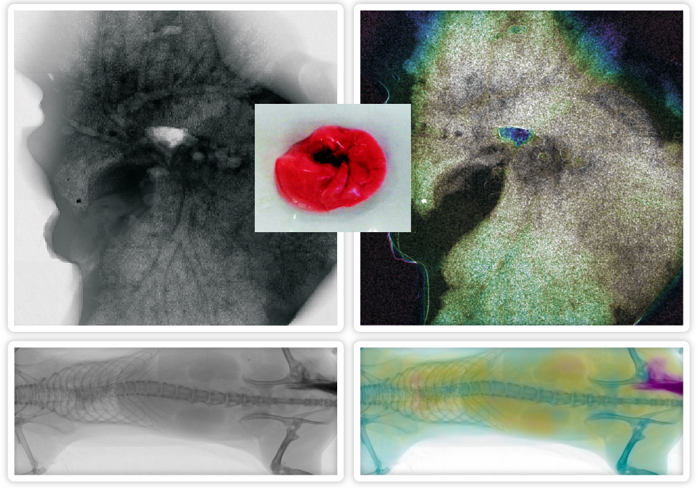
Our devices employ a unique blend of absorption imaging and phase contrast enhancement, allowing clear visualization of specific details in large tissue samples.

We have demonstrated this technology’s power by imaging a deflated mouse lung sample. Traditional imaging techniques struggled to differentiate between the granular structure of alveoli and other lung structures. Our advanced system, however, can distinguish alveoli and different tissue types and structures.

Our high-contrast X-ray system fine-tunes the X-ray beam spectrum and detector energy sensitivity levels to generate detailed images. This technology is a potent tool for tissue analysis, revealing features typically hidden due to low contrast, thereby revolutionizing the study of lung microstructures.

There are two outputs of our single-particle camera. On the left, there's an image showcasing anatomic features: The veins or arteries of mouse lungs clearly. The right image employs a detector to visualize a specific micro-structure: The lung's alveoli, allowing for the observation of their distribution within the organ anatomy. The X-ray imaging system utilized in this study merges a microfocus X-ray tube with a large-area photon counting detector, WidePIX 5X10, having a resolution of 2560 x 1280 pixels with a pitch of 55 µm.
There are two outputs of our single-particle camera. On the left, there’s an image showcasing anatomic features: The veins or arteries of mouse lungs clearly. The right image employs a detector to visualize a specific micro-structure: The lung’s alveoli, allowing for the observation of their distribution within the organ anatomy. The X-ray imaging system utilized in this study merges a microfocus X-ray tube with a large-area photon counting detector, WidePIX 5X10, having a resolution of 2560 x 1280 pixels with a pitch of 55 µm.

Share article