ADVACAM Non-destructive testing: SPECTRAL IMAGING FOR NON-DESTRUCTIVE TESTING. An example of spectral X-ray imaging is the differentiation of materials in electronics. A laptop was scanned using the Widepix MPX3 CdTe detector on the robotic scanner. The resulting scan has 7852x9880 pixels, i.e., 77.6M pixels. Both the grayscale and “color” X-ray images are shown.
Applications

SPECTRAL IMAGING FOR NON-DESTRUCTIVE TESTING

Applications: NDT, non-destructive testing

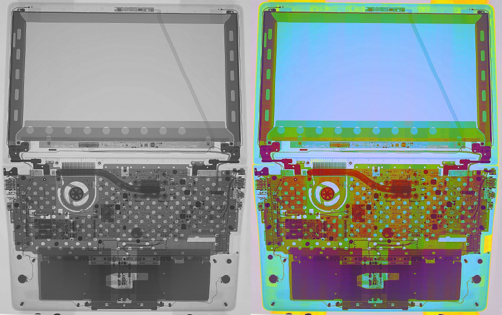
Color X-ray imaging is now achievable. With our spectral detectors, we can measure the wavelength of each X-ray photon and determine the material being scanned. This technology revolutionizes non-destructive testing in electronics and various other complex devices, eliminating the need for disassembly. 

Conventional X-ray images are grayscale, representing only variations in the density and thickness of materials. A thin layer of high-density, heavy material may appear at the same gray level as a thick layer of lightweight material, causing some features of samples to remain undetected. In contrast, energy-sensitive (spectral) X-ray imaging provides information on the elemental composition of the inspected object.

For example, this technique can be employed in non-destructive testing of electronics, where specific colors can distinguish different materials.

ADVACAM Non-destructive testing: SPECTRAL IMAGING FOR NON-DESTRUCTIVE TESTING. An example of spectral X-ray imaging is the differentiation of materials in electronics. A laptop was scanned using the Widepix MPX3 CdTe detector on the robotic scanner. The resulting scan has 7852x9880 pixels, i.e., 77.6M pixels. Both the grayscale and “color” X-ray images are shown.
ADVACAM Non-destructive testing: SPECTRAL IMAGING FOR NON-DESTRUCTIVE TESTING. An example of spectral X-ray imaging is the differentiation of materials in electronics. A laptop was scanned using the Widepix MPX3 CdTe detector on the robotic scanner. The resulting scan has 7852×9880 pixels, i.e., 77.6M pixels. Both the grayscale and “color” X-ray images are shown.


Share article