ThyroPIX – New generation of the camera for the thyroid gland and small organs imaging by nuclear medicine methods.
The project is part of the 1. Public contest for industrial and experimental development program TREND organized by TAČR. It took the 11th place amongst all 396 projects submitted in this contest. The project is scheduled for 4 years, beginning in 2020.
The consortium was established upon the previous long-term cooperation of all the incorporated companies.
Advacam s.r.o. is the leader of the project, having successfully completed several projects in the past – not only in Czech Republic, but on an international scale as well. ADVACAM is responsible for the part dedicated to the development of the detectors and advanced imaging methods.
The other members of the consortium are:
- Radalytica a.s.: integration of the imaging system into the final solution within developing the control software and imaging software
- Czech metrology institute: Monte Carlo simulations and imaging methodology
- Charles University, 1. Medical Faculty, Center of advanced preclinical imaging: preclinical tests on small animals
- University hospital in Motol, Clinic of nuclear medicine and endocrinology: clinical tests on human patients
The main goal of the project is to develop a new mobile robotic imaging camera prototype for thyroid gland and small organ imaging by nuclear medicine methods in diagnostic and therapeutic procedures in human medicine. ThyroPIX will bring an efficient instrument to meet the 2018/02 implemented EU ordinance 2013/59/EURATOM requirements on radiation therapy planning and verification, which is problematical in nuclear medicine methods.
The major concern in design and development will be to eliminate any known limitations and disadvantages, which prevents the therapeutic centers to meet the requirements of the legislations. This will consequently enable the application of principals of personalized medicine in administration of the therapeutic radio-pharmaceuticals.
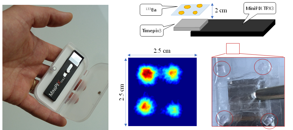
Fig. 1 Detector MiniPIX TPX3 with CdTe sensor of 2 mm (left) and results of “”proof-of-concept” experiment visualizing the distribution of four sources of Ba-133 which has similar energy spectrum as I-131 used in medicine. This test demonstrates the proper function of the Compton Camera including the spatial resolution at the level of millimeters.
The main advantages of this innovative camera ThyroPIX are:
- use o hybrid phonon counting pixels detector allowing single-photon detection of gamma photons in fully-spectral mode (Timepix3 technology)
- High activity radionuclide accumulated in target volume imaging for treatment verification
- Detection of high photon fluxes
- High spatial resolution imaging unavailable with current state-of-art gamma cameras using collimators
- Lowering of diagnostic activities due to high detection efficiency
- Shortening acquisition times due to high detection efficiency
- Combination of planar and tomographic imaging (2D and SPECT) thanks to robotic arm implementation
- Mobile camera concept
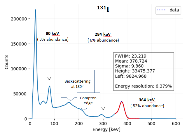
Fig. 2 The energy spectrum of 131I and energy resolution of MiniPIX Timepix3 detector. The spectrum shows even energy peaks at the emission lines with the lower emission.
Strategy of the consortium is the development of advanced imaging system dedicated primarily to thyroid gland monitoring
- Before the administration of therapeutic radioiodine
- provide detailed information about the size a topography of the gland’s remnants
- provide data for the optimal radioiodine activity for those thyroid tissue remnants ablation
- During the treatment
- verify the distribution of therapeutic activity in the target volume
- provide the information about dynamics of accumulation of radioiodine
- and after radioiodine treatment
- to determine the patient’s homecare-release radioiodine activity.
By **final product, our consortium understands the advanced camera should integrate two highly innovative elements::
- Detector in Compton camera configuration based on hybrid pixel detectors technology of physical pixel size of 55 µm
- Collaborative robotic arm which enables as planar as 3D volume reconstruction of data acquired directly on the patient’s bed. Also, eliminate the need for transportation to a dedicated gamma camera inspection room, which minimizes the possible contamination and irradiation of the personnel and other staff.
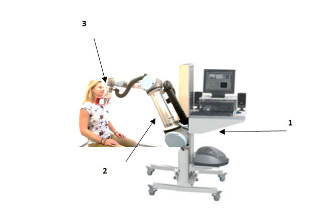
Fig. 3 Illustrative concept of ThyroPIX camera: 1) mobile base with IT and peripheries for robot motion control and acquisition parameters setting. 2) robotic arm Universal Robots UR5 with safety sensors and optical system. 3) hybrid pixel detector for photons detection
ThyroPIX will be equipped also with
- proprietary hardware (control unit) and software for motion control of the robot
- acquisition software for data collection in planar and SPECT modes
- processing software
- software modules for compatibility and communication with systems
- NIS/RIS
- PACS
- suitable hardware (PC) with all the software packages
- equipment for wireless data handling
- and optional upgrade with other software packages
The developed camera will be intended as:
- be dedicatedto diagnostic imaging and imaging of therapeutic activities assuring the measurements in required number and periods.
- offer significantly better resolution
- 2D – <2 mm
- SPECT – <0,5 ml
- substantially shorten acquisition time because of no collimator implementation (especially in SPECT)
- enable detection of high photon fluxes for verification of spatial distribution of therapeutic radiopharmaceuticals
Due to the flexibility and automatic robotic arm positioning, the patient can sit on the chair or lie down in bed without the need of transportation. This helps prevent radioactive contamination. Furthermore, the constant positioning assures the correct evaluation of repeated measurements and the trend curves delineation. The preparation for IIa risk classification healthcare device certification is also part of the project.