FullSpect3D –Fully Spectral Multimodal 3D imaging system for small animals
FullSpect3D -Fully Spectral Multimodal 3D imaging system for small animals FullSpect3D is part of the third public competition in the TRIO program, organized by the Ministry of industry and trade. The project is scheduled for 3 years, beginning in 2018. Advacam s.r.o. is leading the project, having successfully completed several projects in the past – not only in the Czech Republic, but on an international scale as well. ADVACAM is responsible for the part dedicated to the development of the detectors and advanced imaging methods.
The other members of the consortium are:
- Radalytica a.s.: Integration of the imaging system into a final solution and developing the control software and imaging software.
- Charles University, 1. Medical Faculty, Center of advanced preclinical imaging: Evaluation of the developed system and preclinical tests on small animals.
Studies on small animals are a basic and inseparable part of the research for new drugs and methods of cancer treatment. The main aim of this project is the development of a new multimodal 3D imaging system for preclinical and biological studies on small animals, allowing a significant increase of scanning speed, the reduction of radiation dose and improvement of image quality. The system combines three main imaging modalities:
- SPECT (Single Photon Emission Computed Tomography)
- Compton Camera
- PET (Positron Emission Tomography)
- spectral CT (Computed Tomography)
- XRF imaging (X-Ray fluorescence) for visualization of contrast agents or heavy elements naturally occurring in the organism
The detection system exploits the newest technology of Timepix3 detectors which makes it possible to detect individual photons and simultaneously measures their energy, incidence time and position.
Main benefits of this imaging system include:
- reduction of the radiation exposure of the animal during imaging,
- increase in speed of measurement and thus streamlining preclinical studies,
- reduction of contrast and / or radioisotope for functional imaging,
- combining of imaging modalities into one device resulting in
- reduction of scanner acquisition costs.
Other possible application:
- imaging of pathological samples
- imaging of biopsy specimens
- monitoring of therapeutic procedures (brachytherapy, thyroid gland …)
- imaging using mammography
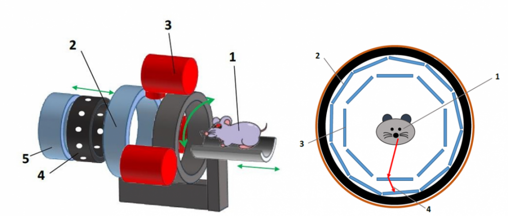
Fig. 1 Fully spectral imaging system: contains: 1. bed with anesthetic support, 2. ring with primary detectors, 3. X-ray tubes, 4. ring with collimators, 5. Optional secondary inner ring with detectors for Compton camera
FullSpect3D system design
The small animal imaging system is designed for rodents such as mice or rats (see Fig 1). The system is as small and compact as possible. Its heart is the stationary cylindrical detector module assembled of 20 Timepix3 detectors with 2 mm thick CdTe sensors. The detector module surrounds the sample. The rotational gantry carries up to three X-ray tubes, serving as source of the primary beam for purposes of transmission x-ray computed radiography and XRF imaging. The shielded circle or semicircle with multi-hole collimators is automatically inserted into the detection module for XRF and SPECT measurements. These components together enable simultaneous or sequential measurements using several imaging modalities.
- Spectral x-ray transmission radiography
- Spectral high resolution, high contrast SPECT
- High resolution PET with high SBR
- Additional modalities such as: Compton camera, XRF imaging, back-scatter imaging …
First prototype
The first prototype served as a system for setting of the geometrical configuration of the final prototype, testing of imaging methods, developing of control software and reconstruction software. This system consists of two X-ray tubes in the opposition and two spectral detectors AdvaPIX TPX3. They are also in the opposite to two Widepix 2×5 imaging detectors. All of the sensors assembled on the cameras are CdTe. The imaging cameras are equipped with a 1mm thicker sensor. The spectral cameras AdvaPIX TPX3 are assembled by a 2mm thicker sensor, due to higher detection efficiency for higher energies (PET).
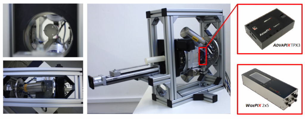
Fig. 2 Mechanical design of first prototype imaging system. Images represent different views on the device.
Circular detector – heart of the system
We say the heart of the system because it is the main part of the imaging platform. The system would not work without it. A single layer of detectors creates the detection unit. Originally, the circular detector should have been created two layers (two-layer Compton camera). The decision to make a circular detector was made when the concept of a single layer Compton camera was successfully proven and tested. The circular detector consists of 20 individual AdvaPIX TPX3 detectors made of a 2 mm thick CdTe sensor. CdTe sensor material was selected based on the results and characteristics of this sensor type. Each detector is connected to electronics using a flexible cable, which allows for the cylindrical shape of this detection unit.
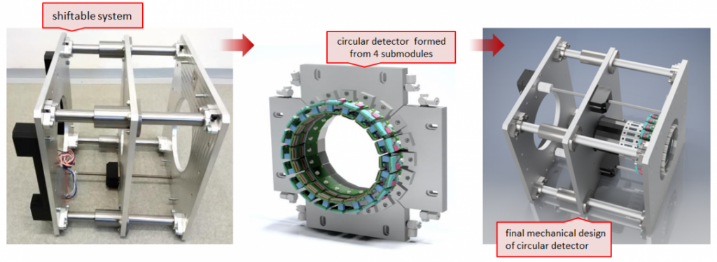
Fig. 3 Circular detector and its components.
The detector is formed by 4 submodules. Each of them is set up to form 5 assemblies. These submodules are then set up together and create the detector of a circular shape with a90 mm radius. This detection unit is mounted on to an adjustable stage, which allows the inserting of a collimation system. The collimation system is made of 20 single pinhole collimators with double conic shape. The collimators are made of wolfram with a thickness of 3 mm. It is sufficient to shield energy up to 150 keV.
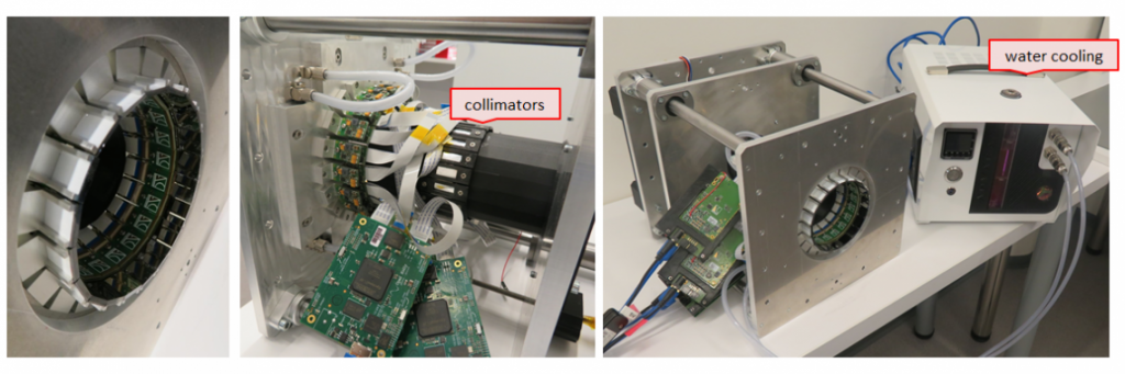
Fig. 4 An constructed circular detector
SPECT/PET/XRF scenner – final prototype
The detection unit (circular detector with the collimation system) was placed on the specially prepared holder, allowing for the x-ray tube to move around the circular detector. The X-ray tube is mounted on to a robotic arm. It allows continual and controlled motion in all of its axis. The use of the X-ray tube, in combination with the robotic arm, was selected due to the impossibility to do a CT scan according to the original configuration. Based on the results using one robotic arm, we decided to implement a second robotic arm. This has made it possible to hold the bed for small animals and to slide it into the detection unit. Thanks to the use of robotic arms, the system becomes modular. Modularity is one of the major benefits of this system. Individual parts of the scanner can be easily dismantled and used for other applications. The scanner can be placed into a shielded box to create a compact imaging system for small animals.
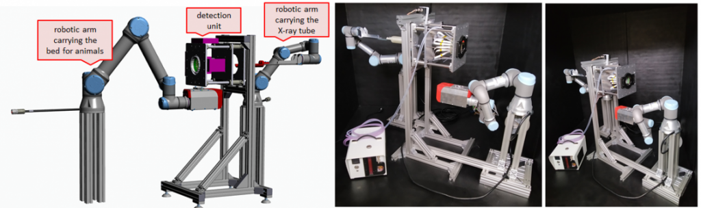
Fig. 5 SPECT/PET/XRF scanner – final prototype.