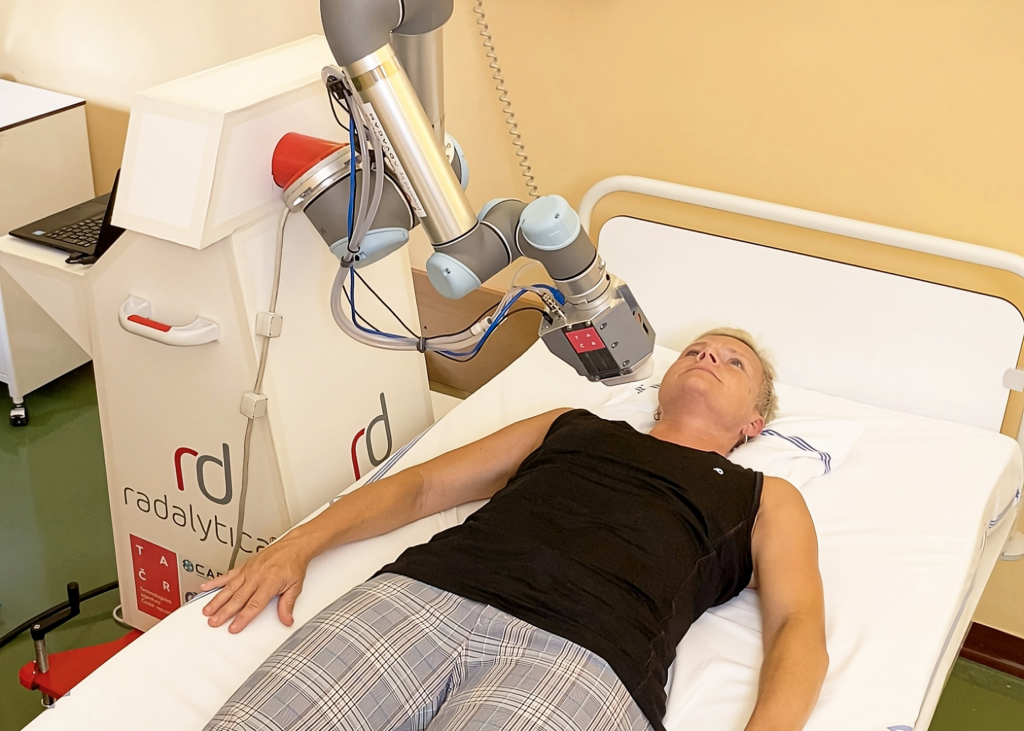
A newly developed robotic device called ThyroPIX could help to more accurately map the distribution of radioactive iodine in the detection and treatment of thyroid tumors. It uses a particle camera to precisely locate where and how the radiopharmaceutical acts.
The aim of the Thyropix project was to develop a unique medical device that will improve the possibilities of monitoring the effect of radiopharmaceuticals and minimize their possible side effects.
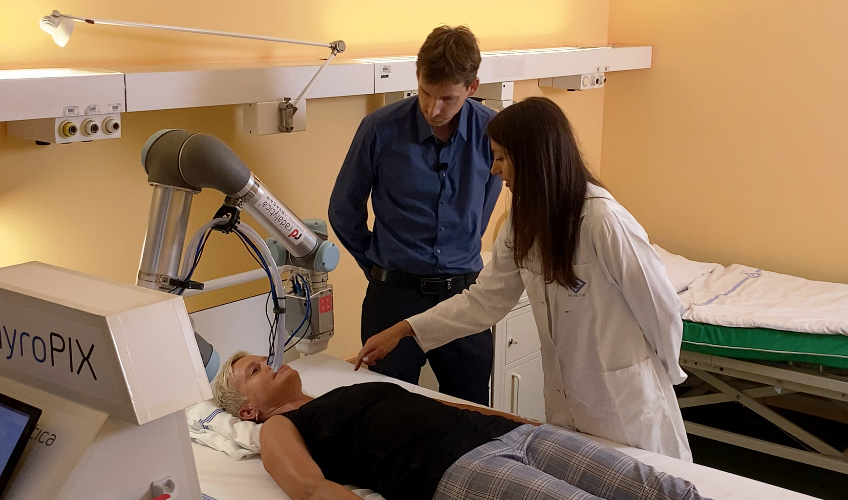
The ThyroPIX uses a robotic arm to get closer to the thyroid and can capture it more accurately and always in the same way during repeated examinations. At the heart of the device are particle cameras manufactured by ADVACAM.
It uses Compton scattering to determine the direction and energy of each individual incoming particle of ionizing radiation. In this way, it is possible to obtain detailed information on the size and shape of thyroid residues, thus verifying the distribution of therapeutic activity in the patient’s body.
ThyroPIX has been tested on a phantom model developed by the Czech Metrology Institute. They also created a complete computer simulation of the entire detection system. “The reason was so that our colleagues from ADVACAM did not have to produce dozens of different combinations of sensors,” explains Jan Rusňák from the Department of Primary Metrology of Ionizing Radiation of CMI. The camera was tested at the Center for Advanced Preclinical Imaging, 1st Faculty of Medicine, Charles University. “The main advantage of ThyroPIX is that it offers standardization of examinations, a wide field of vision and higher sensitivity than other devices,” says Luděk Šefc, head of the center, adding: “The compactness and the associated mobility of the device are also great. Thanks to it, it is possible to examine a patient directly in bed.”The existing methods often cannot sufficiently help in deciding on the most appropriate treatment strategy. “Physically, the devices commonly used today are not able to have such a resolution for iodine-131,” explains Tereza Kráčmerová, a clinical radiological physicist at the University Hospital in Motol. “We see a few spots there, but with poor spatial resolution, we are not able to pinpoint their exact location,” she adds. In addition, an examination takes a long time – about 20 minutes.