Precise X-ray images can detect malignant diseases such as tumours. Many scientists are developing the capabilities offered by subatomic particle detectors in their research. An image taken with a “camera” that can capture individual photons (called photon-counting technology), produced by the Czech company ADVACAM, has made the cover of the scientific journal Journal of Medical Imaging (JMI).
The 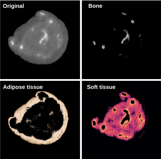 image is in fact a collage of results published in the eleventh issue of JMI by experts from the University of Houston, USA. They are using these “cameras” to push the limits of computed tomography. Their aim is to achieve the most accurate results in distinguishing different types of biological tissue. This can be useful for detecting unwanted growths and tumours or observing areas that conventional techniques cannot capture.
The Houston researchers looked at ways to improve imaging results by using photon-counting detectors to image and distinguish biological materials in the body. As part of a collaborative project, ADVACAM also developed a unique model of the WidePIX large-area detector for the University of Houston that can image an area of 1,638,400 pixels.
The photon-counting detector technology that ADVACAM produces was primarily developed for experimental purposes related to matter research at the Large Hadron Collider (LHC) at CERN. These detectors make it possible to obtain important information about each particle that comes into contact with their sensor, offering very high resolution imaging and the ability to discriminate between materials. They can be used for X-ray imaging in biomedicine, non-destructive testing, particle physics research or monitoring radiation in space.