By Daniel Turecek
Positron emission tomography (PET) is a nuclear medicine functional imaging technique. It is used in clinical oncology (medical imaging of tumors and the search for metastases) and pre-clinical studies using animals. PET uses small amounts of radioactive materials (radiotracers) and a special photon sensitive camera. Most of these cameras use scintillators with photomultipliers as detectors. However, these detectors have limited energy sensitivity and large pixels. The false signal caused by a scattering poses a significant problem. However, Science Direct presents an initial study and evaluation of two Timepix3 detectors with 2 mm thick CdTe sensors used in simplified geometry for PET imaging. The study is performed on 2 samples — a capillary tube and a cylindrical plexiglass phantom with cavities. Both samples are filled with fluodeoxyglucose (FDG) solution that is used as a radiotracer. The Timepix3 offers better properties compared to conventional detectors — high granularity (pixel pitch), good energy resolution (1 keV at 60 keV) and sufficient time resolution (1.6 ns). The spectral sensitivity of Timepix3 together with coincidence/anticoincidence technique allows for significant reduction of background signal caused by compton scattering and internal X-ray fluorescence of Cd and Te.
The precision of the time measurement achieved with Timepix3 is sufficient for identification of coincidence events. The Timepix3 spectro-scopic properties allow separation or in some cases even reconstruction of the valid signal out of different types of background events, such as different types of Compton scattering, internal fluorescence of CdTe sensors and their combinations. This feature substantially decreases the amount of false information acquired during PET measurement and greatly improves the signal to background ratio of the reconstructed images (by two orders of magnitude). It presents a great advantage, especially in the field of small animal imaging, where the traditional method of background suppression based on the time-of-flight technique does not reach sufficient resolution due to the small sample size.
This study shows that the Timepix3 detector with CdTe sensors are significantly more efficient in PET imaging of small animals. Although the measurements and reconstructions in this work were performed only in 2D, the same principles can be applied for the 3D reconstruction (using multiple detectors surrounding the measured sample). The full 3D reconstruction will also be evaluated in future work.
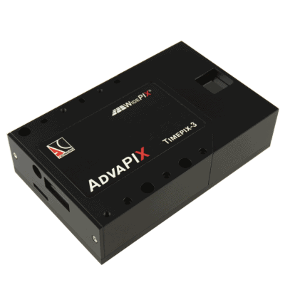
Moreover, the compact size, portability and USB3 connectivity of Timepix3 data acquisition system has a great advantage, enabling construction of a very compact and portable PET system for preclinical tests with small animals such as mice or other rodents.
The Timepix3 technology could also be used in the future for patient’s PET scanners. The greatest benefit is the high selectivity of the detection that enables us to visualize even very small structures in proximity of large structures (cancer remnants after surgery). These structures would be concealed by background signal. The other benefit of using Timepix3 would be possible reduction of the dose, but further investigation is needed.