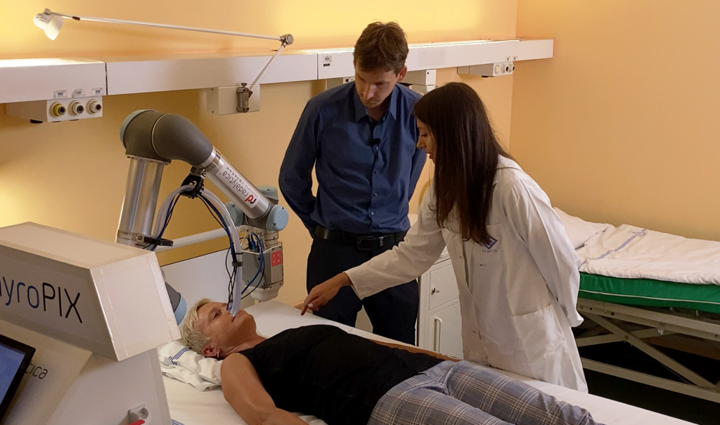
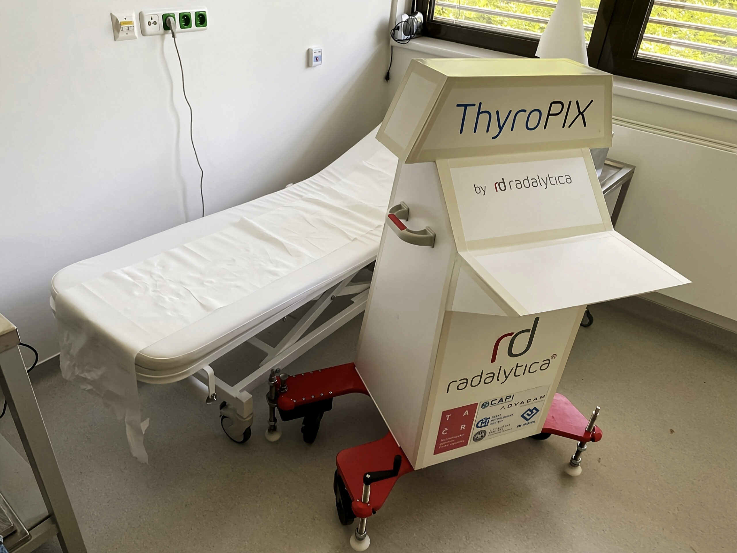
The newly developed robotic device could help to more accurately map the distribution of radioactive iodine in the detection and treatment of thyroid tumors. Thanks to the groundbreaking miniaturized gamma camera, capable of determining the direction of incoming radiation, it precisely locates where and how the radiopharmaceutical acts. The prototype, called ThyroPIX, was developed in as part of the project of the same name financed by the Technology Agency of the Czech Republic.
Doctors worldwide diagnose approximately 300,000 new cases of thyroid cancer every year. One common part of the treatment is radioiodine therapy, which is usually performed after surgical removal of the tumor. Even after surgery, there are usually tiny remnants of tumor tissue left in the patient’s throat and these need to be removed to prevent the disease from returning. Patients therefore receive a radioactive isotope of iodine, which accumulates naturally in the thyroid gland and irradiates the affected area locally, thus eliminating the cancerous growth.
The aim of the Thyropix project was to develop a unique medical device that will improve the possibilities of monitoring the effect of radiopharmaceuticals and minimize their possible side effects. Members of the consortium, which was supported in this successful research by the Technology Agency of the Czech Republic, were the 1st Faculty of Medicine of Charles University, Motol University Hospital, the Czech Metrology Institute and innovative companies Radalytica and ADVACAM.
The aim is to overcome the physical limits of existing methods
The existing methods often cannot sufficiently help in deciding on the most appropriate treatment strategy. “Physically, the devices commonly used today are not able to have such a resolution for iodine-131,” explains Tereza Kráčmerová, a clinical radiological physicist at the University Hospital in Motol.
“We see a few spots there, but with poor spatial resolution, we are not able to pinpoint their exact location,” she adds. In addition, an examination takes a long time – about 20 minutes.
ThyroPIX uses a robotic arm to get closer to the scanned area and can capture it more accurately and always in the same way during repeated examinations. At the heart of the device are particle cameras manufactured by ADVACAM. Thanks to a newly developed methodology for using the so-called Compton scattering, it can determine the direction and energy of each individual incoming particle of ionizing radiation. In this way, it is possible to obtain detailed information on the size and shape of thyroid residues, thus verifying the distribution of therapeutic activity in the patient’s body.
Software creates a 3D image of the distribution of radioactive iodine in the patient’s body
The software that the scientists developed as part of the project is crucial for doctors – only in the computer does the data obtained turn into an image with visible remains of the tumor. The gamma camera sensor captures the scattering of charge that occurs when a photon passes through two layers of the sensor with different properties.
“A photon flies out of the primary radiation source, which hits the first layer of sensitive material, where part of the energy is transferred, and a Comptonian photon flies out from the same place, and interacts in the second layer,” says the project’s investigator Eliška Trojanová, describing the principle of the detector. The device then calculates the direction from which the radiation comes and with enough photons it determines the exact location of the radiation source.
ThyroPIX has been tested on a phantom model developed by the Czech Metrology Institute. They also created a complete computer simulation of the entire detection system. “The reason was so that our colleagues from ADVACAM did not have to produce dozens of different combinations of sensors,” explains Jan Rusňák from the Department of Primary Metrology of Ionizing Radiation of CMI.
The camera was tested at the Center for Advanced Preclinical Imaging, 1st Faculty of Medicine, Charles University. “The main advantage of ThyroPIX is that it offers standardization of examinations, a wide field of vision and higher sensitivity than other devices,” says Luděk Šefc, head of the center, adding: “The compactness and the associated mobility of the device are also great. Thanks to it, it is possible to examine a patient directly in bed.”
When will ThyroPIX reach real patients?
However, there are still a few steps left to put the innovation into practice. The gamma camera head needs to be adjusted to get even closer to the thyroid gland. Further software improvements are planned so that the device can eventually undergo clinical trials directly on patients. The authors are also now intensively looking for an industrial partner that would be interested in helping to bring the solution to the market as a finished product.
“We would prefer to hand over this entire solution to someone who already has experience with medical device development and the certification process,” concludes the project’s principal investigator from ADVACAM, Eliška Trojanová.
This project is funded with state support from the Technology Agency of the Czech Republic and the Ministry of Industry and Trade of the Czech Republic under the TREND Programme