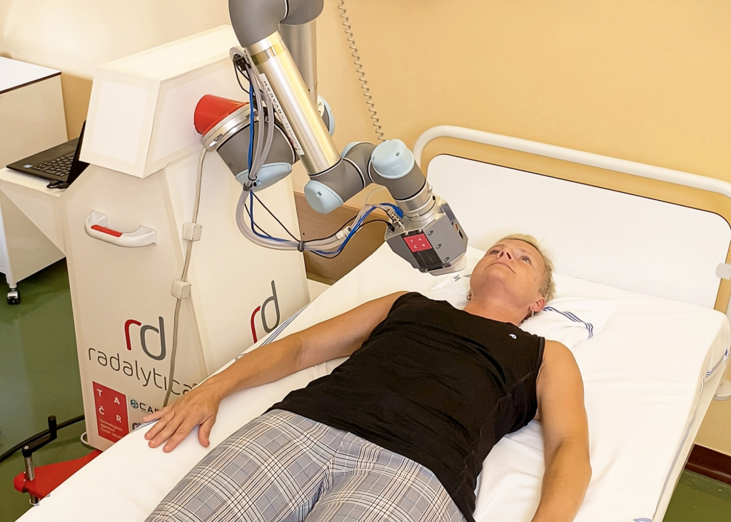
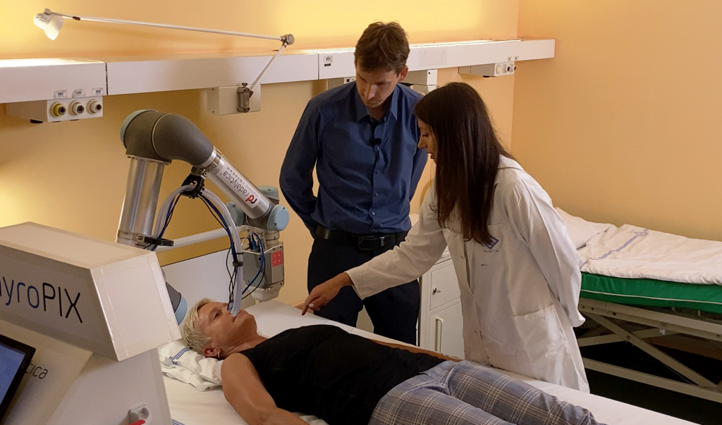
ROBOT MONITORS RADIOACTIVE DRUGS IN THE BODY
A newly developed robotic device called ThyroPIX could help to more accurately map the distribution of radioactive iodine in the detection and treatment of thyroid tumors. It uses a particle camera to precisely locate where and how the radiopharmaceutical acts.
The aim of the Thyropix project was to develop a unique medical device that will improve the possibilities of monitoring the effect of radiopharmaceuticals and minimize their possible side effects.
The ThyroPIX uses a robotic arm to get closer to the thyroid and can capture it more accurately and always in the same way during repeated examinations. At the heart of the device are particle cameras manufactured by ADVACAM.
It uses Compton scattering to determine the direction and energy of each individual incoming particle of ionizing radiation. In this way, it is possible to obtain detailed information on the size and shape of thyroid residues, thus verifying the distribution of therapeutic activity in the patient’s body.
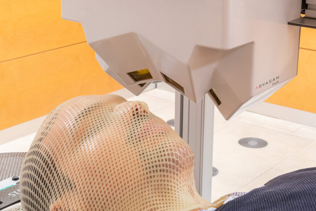
MORE PRECISE HEAD CANCER TUMOR IRRADIATION
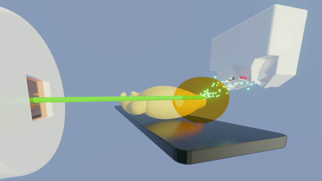
ADVACAM detectors are being tested as a method to improve head cancer ion beam irradiation. The new device Beam TraX allows treatment of a smaller tissue volume, which can help reduce negative side effects.
The tests focus on patients with tumors near the base of the skull. This area is challenging to access for irradiation due to the proximity of essential structures like the brainstem. This device called Beam TraX measures secondary radiation emitted from the patient, and based on this data, scientists understand what kind of matter is being irradiated.
So far, doctors had to rely on previously done CT scans. However, the situation inside the patient’s head can change during the therapy. The new device is meant to allow a better understanding of where and how often these changes occur. With its use, doctors can save healthy tissue from irradiation and apply higher doses to the tumor.
The presence of detectors does not affect the existing therapy. It can help prevent side effects such as memory or optic nerve damage.
The InViMo clinical trials are being conducted by scientists from the German National Center for Tumor Diseases (NCT), the German Cancer Research Center (DKFZ), and the Heidelberg Ion Beam Therapy Center (HIT) at Heidelberg University Hospital.
SPECTRAL RADIOGRAPHY
Photon counting detectors excel in bio-imaging due to their high sensitivity and dynamic range.
The high sensitivity of photon counting detectors makes them helpful in imaging low X-ray attenuating light objects, such as tissue. Thus, these detectors are ideal for bio-related applications. High dynamic range reveals features in samples that remain hidden from other types of X-ray imaging detectors. Moreover, the structure of the model can be visualized thanks to the spectral sensitivity of the device. Only specific structures can be highlighted, and other ones can be suppressed.
SPECTRAL COMPUTED TOMOGRAPHY
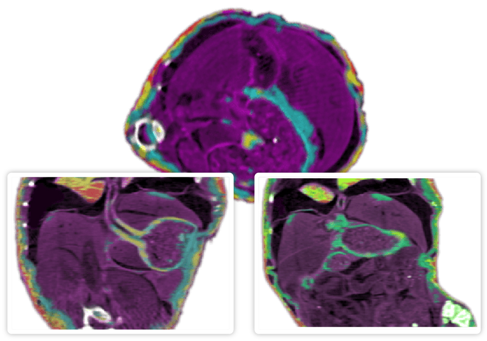
Spectral computed tomography enhances 3D imaging, advancing tissue identification and cancer treatment research.
Spectral radiography can be extended to 3D using computed tomography. This makes it possible to recognize different types of tissue in natural form. This level of information can be beneficial for cancer treatment research, as it gives better data for irradiation planning.
SOFT TISSUES DIFFERENTIATION
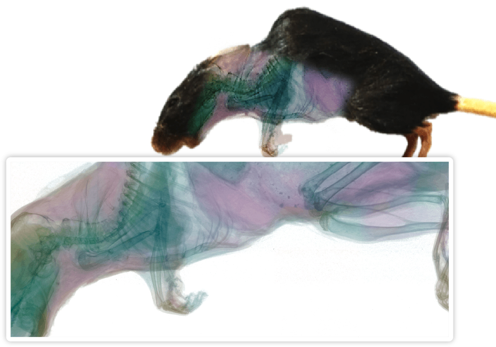
Our innovative imaging cameras employ advanced technology for enhanced clarity and detail in extensive tissue sample analysis.
Our devices employ a unique blend of absorption imaging and phase contrast enhancement, allowing clear visualization of specific details in large tissue samples.
We have demonstrated this technology’s power by imaging a deflated mouse lung sample. Traditional imaging techniques struggled to differentiate between the granular structure of alveoli and other lung structures. Our advanced system, however, can distinguish alveoli and different tissue types and structures.
Our high-contrast X-ray system fine-tunes the X-ray beam spectrum and detector energy sensitivity levels to generate detailed images. This technology is a potent tool for tissue analysis, revealing features typically hidden due to low contrast, thereby revolutionizing the study of lung microstructures.
FUNCTIONAL REAL-TIME TISSUE IMAGING
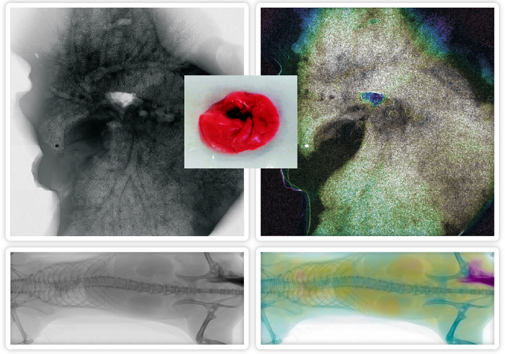
Our cameras provide real-time, high-speed X-ray videos of live inner organs, revealing intricate details like a rodent’s heartbeat.
Leveraging the unique capabilities of our advanced imaging cameras, we offer real-time visualization of dynamic processes within organs and tissues. Our cameras’ exceptional speed enables capturing thousands of X-ray-sensitive images per second, creating high-speed X-ray videos of live inner organs and tissues.
This technology allows for incredible detail, such as observing a rodent’s heart’s blood flow. Additionally, our method can monitor the movement of contrast agents within the body in real-time, providing invaluable insights into dynamic organ functions, including the kidneys, brain, muscles, and joints.
This footage shows a slow-motion recording of a mouse’s heartbeat. The rodent’s heart beats at a rate of 670 beats per minute, ten times faster than a human heart. The recording was made with our AdvaPIX detector at a rate of 1000 frames per second. Bone tissue was suppressed using post-processing to make the heart visible even behind the rib cage. In the video, you can observe how the individual chambers and atria of the heart darken or lighten, which is the actual movement of blood inside the organ. In this case, no contrast agent was used. Go on AdvaPIX models ›

FOOD INSPECTION: X-RAY CLASSIFICATION AND QUALITY CONTROL
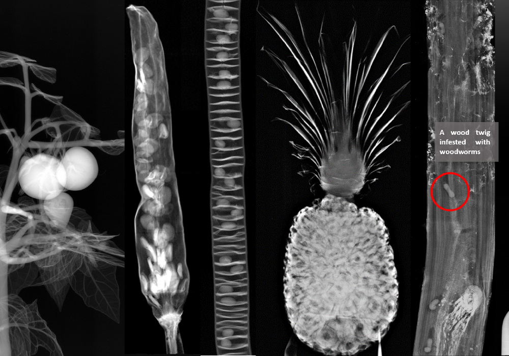
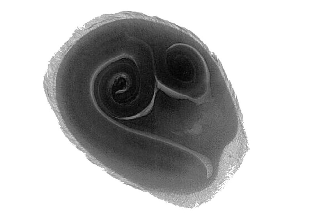
Our single-photon-counting cameras allow for the creation of detailed X-ray material-sensitive images that reveal the internal composition of various food items, including fruits, vegetables, and grains.
ADVACAM‘s technology enables fast real-time inspection of qualities typically hidden to the naked eye. For example, sprouts – an indicator of potential growth – can be clearly identified in a detailed X-ray of a tomato seed. If these sprouts are absent, it suggests that the seed is unlikely to germinate.
Our imaging technology also allows for early detection and characterization of crop yield. For instance, as a cereal plant develops, X-ray imaging can reveal the ear grains even when they are still growing through the stem and not visible externally. Individual ears and the prospective number of grains can be predicted, allowing for early yield estimation – a critical factor in plant breeding.
It is possible to view various types of food through their packaging using non-destructive testing and thus monitor quality.
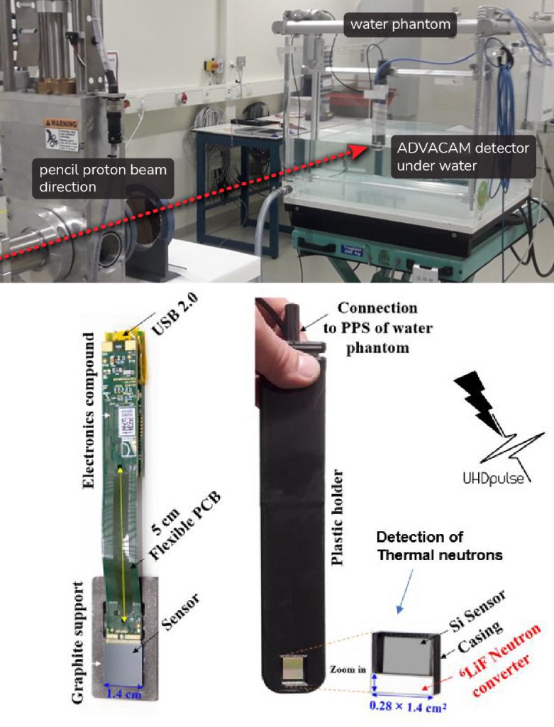
MINIMIZING SIDE EFFECTS OF INNOVATIVE CANCER RADIOTHERAPY
Our single-photon counting detectors participated in a groundbreaking research project on ultra-high pulse dose rate cancer radiotherapy. This innovative approach might dramatically reduce side effects while maintaining effective tumor control.
The World Health Organization estimates 4.2 million new cancer cases in Europe alone in 2018, with approximately half of the patients receiving radiotherapy. The so-called FLASH effect, observed when radiation doses are delivered quickly via a few ultra-high dose pulses, suggests the potential for more effective tumor control.
However, accurate dosimetry is crucial to ensure safety and effectiveness in radiotherapy, and this is where ADVACAM’s expertise plays an important role. We are focusing on developing methods for characterizing stray radiation, which could contribute to parasitic doses to healthy tissues outside the target volumes. Accurate measurement of this radiation is vital for therapy optimization and personalized dose management.
The research was conducted within the UHDpuse project in cooperation with prestigious international partners.