There are two outputs of our single-particle camera. On the left, there's an image showcasing anatomic features: The veins or arteries of mouse lungs clearly. The right image employs a detector to visualize a specific micro-structure: The lung's alveoli, allowing for the observation of their distribution within the organ anatomy. The X-ray imaging system utilized in this study merges a microfocus X-ray tube with a large-area photon counting detector, WidePIX 5X10, having a resolution of 2560 x 1280 pixels with a pitch of 55 µm.
Advacam WidePIX L 2x15

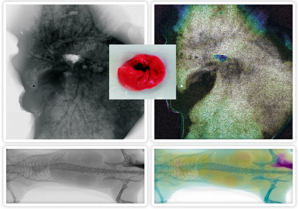
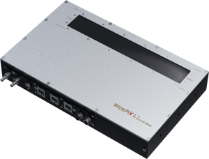
There are two outputs of our single-particle camera. On the left, there’s an image showcasing anatomic features: The veins or arteries of mouse lungs clearly. The right image employs a detector to visualize a specific micro-structure: The lung’s alveoli, allowing for the observation of their distribution within the organ anatomy. The X-ray imaging system utilized in this study merges a microfocus X-ray tube with a large-area photon counting detector, WidePIX 5X10, having a resolution of 2560 x 1280 pixels with a pitch of 55 µm.